





Expo
view channel
view channel
view channel
view channel
view channel
view channel
view channel
view channel
view channel
Molecular DiagnosticsHematologyImmunologyMicrobiologyPathologyTechnologyIndustry
Events
Webinars

- Blood Biomarker May Track Bone Metastases in Medullary Thyroid Cancer
- Blood Biomarkers Help Assess Growth Risks During Steroid Treatment in Duchenne Muscular Dystrophy
- New Blood Biomarker Predicts May Predict Preeclampsia and Fetal Growth Restriction
- Biomarker Score Helps Differentiate Takotsubo Syndrome from Myocardial Infarction
- New Biomarker Recommendations Aim to Personalize Stroke Rehabilitation
- Common Genetic Marker Predicts Faster Motor Decline in Parkinson’s Disease
- Blood Test Helps Clarify Prostate Cancer Risk After Elevated PSA
- Secure Cloud-Connected qPCR System Enables Remote Infectious Disease Surveillance
- Project to Develop Maternal Blood Test for Rapid Fetal Oxygen Deprivation Detection
- DPYD Genotyping Assay Supports Safer Fluoropyrimidine Chemotherapy Dosing
- Biomarker-Guided Framework Aims to Guide Mantle Cell Lymphoma Therapy
- New Genetic Findings Reveal Cause of Bone Marrow Failure Syndrome
- Ultra-Portable Device Enables Finger-Prick Blood Testing at Home
- Age-Specific CBC Reference Intervals Support Pediatric Diagnosis in Vietnam
- Spectral Flow Cytometry Assay Enhances MRD Detection in Multiple Myeloma
- Temporal Immune Profiling Reveals How Sepsis States Change Over Time
- Study Identifies Immune Cells That Drive Harmful Autoantibody Responses in COVID-19
- Gut “Memory” May Explain Why IBD Flares Return
- AI-Based Antibody Profiling Predicts Strength of COVID-19 Vaccine Response
- Immune Biomarkers May Predict Recurrent Checkpoint Inhibitor Arthritis
- Gut Microbiome Classifier Improves Colorectal Cancer Risk Stratification
- New Diagnostic Workflow Identifies Bloodstream Pathogens and Antibiotic Response in Hours
- Genetic Marker Identifies Emerging Resistance to Frontline Malaria Drugs
- Surveillance and Susceptibility Testing Track Rising Candida Auris in U.S.
- Plasma Cell-Free DNA Test Enables Earlier Diagnosis of Invasive Fungal Infections
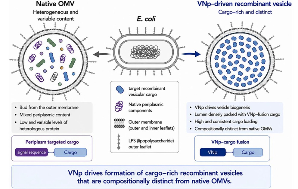
- Bacterial Vesicle Expression System Streamlines Production of Cancer Diagnostic Proteins
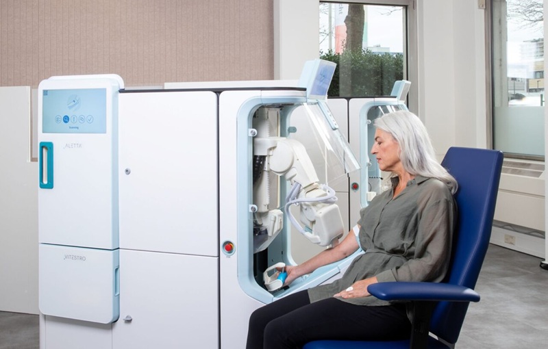
- Autonomous Robotic System Receives FDA Authorization for Blood Collection
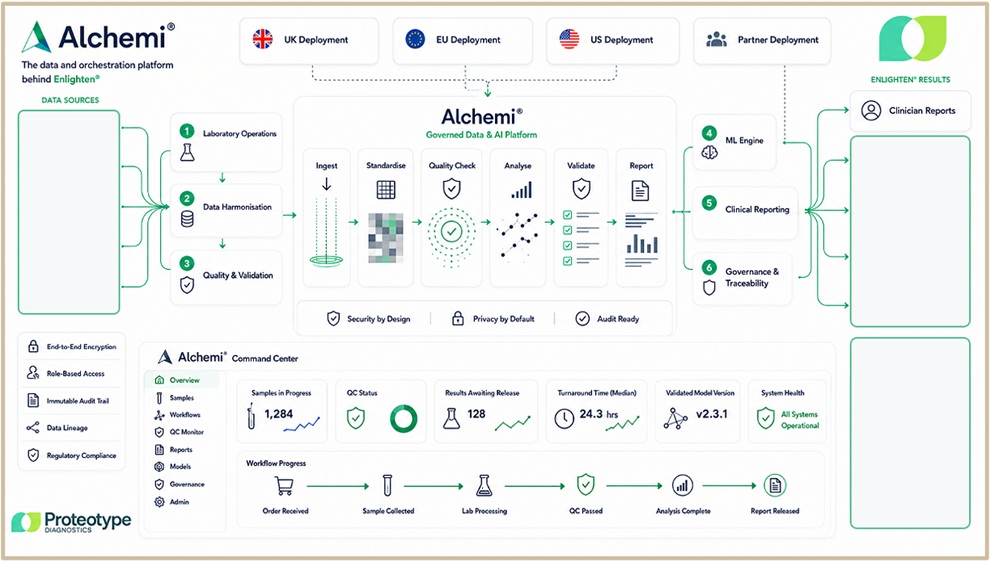
- Interoperable Data Platform Standardizes Multi-Cancer Blood Test Results
- Training Device Improves Accuracy of Pooled Molecular Diagnostics
- New CE-Certified Software Advances Whole-Genome Cancer Testing
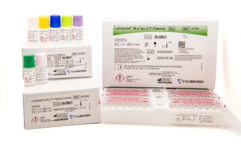
- Fujirebio Expands Access to Automated Blood-Based Neurology Testing in Canada
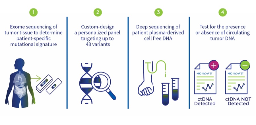
- Natera ctDNA Test to Support Response Monitoring in Solid Tumor Trial
- Tumor-Informed MRD Assay Gains Medicare Coverage for Immunotherapy Monitoring
- Companion Diagnostics Expand HER2 Testing in Metastatic Gastroesophageal Cancer
- Global Urinalysis Market Gowth Driven by Demand for Automated and POC Testing
- New Gene-Disease Link May Help Diagnose Rare Neurodevelopmental Disorders
- Researchers Identify Shared Molecular Networks Behind Fatigue-Related Illnesses
- New Immune Target Could Support More Precise High Blood Pressure Treatment
- Whole-Blood Extracellular Vesicle Analysis Captures Molecular Signals Missed by Plasma Testing
- Blood Metabolite Signature Predicts ALS Progression and Points to Treatment Strategy
- FDA Clearance Expands Digital Pathology Options for Clinical Laboratories
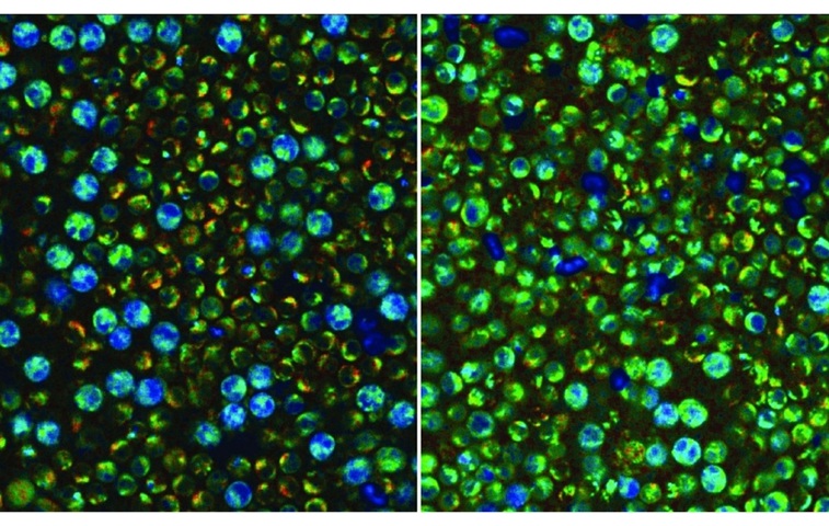
- Label-Free Imaging Reveals Hidden Immune Activity in Blood Samples
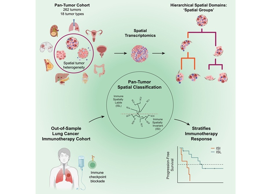
- AI Tool Improves Prediction of Immunotherapy Response in Lung Cancer
- New Journal Addresses Evolving Needs in Clinical and Translational Pathology
- AI Matches Pathologists in Predicting Breast Cancer Prognosis from Immune Cells
 Expo
Expo
- Blood Biomarker May Track Bone Metastases in Medullary Thyroid Cancer
- Blood Biomarkers Help Assess Growth Risks During Steroid Treatment in Duchenne Muscular Dystrophy
- New Blood Biomarker Predicts May Predict Preeclampsia and Fetal Growth Restriction
- Biomarker Score Helps Differentiate Takotsubo Syndrome from Myocardial Infarction
- New Biomarker Recommendations Aim to Personalize Stroke Rehabilitation
- Common Genetic Marker Predicts Faster Motor Decline in Parkinson’s Disease
- Blood Test Helps Clarify Prostate Cancer Risk After Elevated PSA
- Secure Cloud-Connected qPCR System Enables Remote Infectious Disease Surveillance
- Project to Develop Maternal Blood Test for Rapid Fetal Oxygen Deprivation Detection
- DPYD Genotyping Assay Supports Safer Fluoropyrimidine Chemotherapy Dosing
- Biomarker-Guided Framework Aims to Guide Mantle Cell Lymphoma Therapy
- New Genetic Findings Reveal Cause of Bone Marrow Failure Syndrome
- Ultra-Portable Device Enables Finger-Prick Blood Testing at Home
- Age-Specific CBC Reference Intervals Support Pediatric Diagnosis in Vietnam
- Spectral Flow Cytometry Assay Enhances MRD Detection in Multiple Myeloma
- Temporal Immune Profiling Reveals How Sepsis States Change Over Time
- Study Identifies Immune Cells That Drive Harmful Autoantibody Responses in COVID-19
- Gut “Memory” May Explain Why IBD Flares Return
- AI-Based Antibody Profiling Predicts Strength of COVID-19 Vaccine Response
- Immune Biomarkers May Predict Recurrent Checkpoint Inhibitor Arthritis
- Gut Microbiome Classifier Improves Colorectal Cancer Risk Stratification
- New Diagnostic Workflow Identifies Bloodstream Pathogens and Antibiotic Response in Hours
- Genetic Marker Identifies Emerging Resistance to Frontline Malaria Drugs
- Surveillance and Susceptibility Testing Track Rising Candida Auris in U.S.
- Plasma Cell-Free DNA Test Enables Earlier Diagnosis of Invasive Fungal Infections
- Bacterial Vesicle Expression System Streamlines Production of Cancer Diagnostic Proteins
- Autonomous Robotic System Receives FDA Authorization for Blood Collection
- Interoperable Data Platform Standardizes Multi-Cancer Blood Test Results
- Training Device Improves Accuracy of Pooled Molecular Diagnostics
- New CE-Certified Software Advances Whole-Genome Cancer Testing
- Fujirebio Expands Access to Automated Blood-Based Neurology Testing in Canada
- Natera ctDNA Test to Support Response Monitoring in Solid Tumor Trial
- Tumor-Informed MRD Assay Gains Medicare Coverage for Immunotherapy Monitoring
- Companion Diagnostics Expand HER2 Testing in Metastatic Gastroesophageal Cancer
- Global Urinalysis Market Gowth Driven by Demand for Automated and POC Testing
- New Gene-Disease Link May Help Diagnose Rare Neurodevelopmental Disorders
- Researchers Identify Shared Molecular Networks Behind Fatigue-Related Illnesses
- New Immune Target Could Support More Precise High Blood Pressure Treatment
- Whole-Blood Extracellular Vesicle Analysis Captures Molecular Signals Missed by Plasma Testing
- Blood Metabolite Signature Predicts ALS Progression and Points to Treatment Strategy
- FDA Clearance Expands Digital Pathology Options for Clinical Laboratories
- Label-Free Imaging Reveals Hidden Immune Activity in Blood Samples
- AI Tool Improves Prediction of Immunotherapy Response in Lung Cancer
- New Journal Addresses Evolving Needs in Clinical and Translational Pathology
- AI Matches Pathologists in Predicting Breast Cancer Prognosis from Immune Cells


































 Client Login